Cortical areas are areas of the brain located in the cerebral cortex.
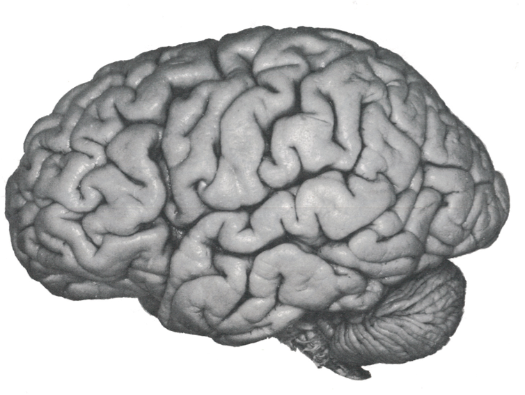
The cerebral cortex refers to the superficial part of the brain and containing the gray matter of the cerebral hemispheres.
The experiments of electrical stimulation and ablation of the cortex made it possible to:
- map the cerebral cortex. This mapping was updated thanks to the technique of cerebral imaging ;
- and thus to locate the cortical areas and determine their functions.
The three types of cortical areas:
Motor areas (or motor cortex)
These areas include the primary motor cortex, premotor cortex, and Broca’s area. Electrical stimulation of these areas causes movements of specific parts of the body.
The primary motor cortex, located in front of the central cleft, controls specific muscles in the body, especially those that cause fine movements (fingers, thumb, lips, mouth).
The premotor cortex, located in front of the motor cortex, causes coordinated movements (e.g. movements engaged in a sporting activity).
Broca’s area coordinates the movements of the larynx and the mouth generating the expression of words. This area is the center of language.
The somesthetic sensory areas (or sensory cortex)
It is a part of the cerebral cortex that is responsible for receiving and interpreting sensory information from different parts of the body: touch, pressure, temperature and pain. The sensory areas occupy the entire temporal lobe.
This area is divided into a primary area and a secondary area. The primary area recognizes particular types of sensations originating from different regions of the body. The secondary area for its essential role is to interpret the signal coming from the body, for example the object that your hand touches is it a chair, a fabric etc.?
The secondary area therefore refines in a way the message received by the primary area.
The primary sensory area is the part of the cortex that directly receives signals from different receptors (such as pain or temperature sensitive receptors). These signals produce an action potential which is transmitted along one or more neurons to a specific part of the brain.
Signals going to the secondary sensory area are processed by the subcortical areas (e.g., the thalamus) or the primary somesthetic area.
- The visual area. The visual area is located in the occipital lobe. The primary visual area specifically detects dark and bright spots, as well as the edge of the visual scene. The role of the secondary visual area is to interpret visual information.
- The auditory area. It is located mainly in the temporal lobe. The primary hearing area perceives specific sounds and sonority. The secondary auditory area interprets the meaning of spoken words.
- The primary olfactory area. It can be found in the front portion of the temporal lobe. It receives input from the olfactory bulb, where signals from olfactory receptor neurons in the nose are processed. This region is essential for distinguishing and recognizing different scents. Interestingly, unlike other senses, olfactory information goes straight to the cortex without passing through the thalamus. Additionally, the olfactory cortex is linked to brain areas associated with emotion and memory, including the amygdala and hippocampus. As a result, certain smells can elicit strong emotional reactions and trigger memories. The areas are the oldest and most conservative area of the telencephalon, play a significant role in various neurologic disorders and neurodegenerative diseases. The recent focus on human olfaction can also be attributed to transitional anosmia caused by COVID-19.
- The primary taste area. It is found in the insular cortex and frontal operculum, and responsible for interpreting taste. Taste buds contain receptor cells that recognize chemical substances from food. These signals are then sent through cranial nerves to the brainstem, thalamus, and ultimately reach the gustatory cortex. Here, the primary tastes of sweet, sour, salty, bitter, and umami are processed. Higher brain areas combine taste with other sensory inputs like smell and texture to form our perception of flavor. The taste and smell systems work hand in hand and provide the brain with information about the chemical composition of objects.
- The somesthetic area
Cortical areas and their functions
Four lobes are used to designate specific anatomical locations and functions of the sensory and motor cortical areas. These regions deal with different modalities of sensation, often relayed by the spinal cord or directly by the cranial nerves. Moreover, one of these regions can even initiate conscious motor movement.
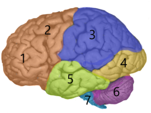
1 and 2. Frontal lobe formed by the prefrontal area (1) and the motor area (2).
The frontal lobe extends from the back of the forehead to the parietal lobe (3). The groove in the cortex known as the central sulcus marks the boundary between the frontal and parietal lobes. The prefrontal area (1) is the control center for executive functions, including reasoning, decision-making, higher level cognitive processes, orientation (person, place, time and situational integration of sensory information). The motor area (2) controls the fine muscles of the body (fingers, lips, mouth), coordinates movement and controls speech (articulation of words).
3. Parietal lobe. Above the temporal lobe and adjacent to the occipital lobe, the parietal lobe houses the primary and secondary sensory cortex. It plays an important role in space navigation and the treatment of touch, pressure, temperature and pain.
4. Occipital lobe. It controls the primary visual cortex, the region of the brain responsible for processing and interpreting visual information. It is located at the back of the brain.
5. Temporal lobe. The temporal lobe extends from the temple to the occipital lobe. It includes the auditory area (detection of auditory signals), Wernicke’s area (interpretation of the meaning of sentences read and heard) and the hippocampus, a structure involved in the formation of memory and emotion.
The other non-cortical structures are the :
6. Cerebellum
7. Brainstem
What are the consequences of a lesion of a cortical area?
A stroke (or stroke) caused by the occlusion of a cerebral artery causes symptoms, the nature and severity vary depending on the cause of the stroke, the region and the extent of damage.
These clinical manifestations are reflected in the symptoms summarized below:
- Difficulty concentrating and planning
- Personality changes (apathy)
- Difficulty performing sequential and / or simultaneous tasks
- Numbness or even paralysis of the body (face, arm, leg) on the side opposite the affected area (total or partial hemiplegia)
- Difficulty or inability to perform actions. The apraxia can be the result of a lesion of the frontal lobes and / or parietal.
- Difficulty speaking (dysarthria)
- Loss of the ability to recognize an object by touch (tactile agnosia) or a person by sight (visual agnosia). Tactile agnosia is observed in lesions of the parietal lobe. The visual agnosia is usually associated with a violation of both occipital lobes. Visual acuity and intellectual functions remain intact.
- Impairment or loss of sight (homonymous hemianospia). A person whose left visual area (i.e. the visual area of the left hemisphere) is affected cannot see objects on the right side of their eyes.
- Speech and thought disorders called Wernicke’s aphasia (the person cannot translate the words read or heard into a coherent thought; sometimes they express themselves inconsistently)
- Short-term memory impairment (loss of the ability to create new memories)
The area of short-term memory (located in each hemisphere) is particularly affected in people with Alzheimer’s disease or mild cognitive decline and, to a lesser extent, in people with stroke (there are compensation phenomena because stroke generally only affects one of the two areas).