Neurons (or nerve cells), fundamental units of the brain and nervous system, have different types and morphologies.
Neurons are the basis of the functioning of the brain. They are surrounded by non-neuronal cells called
glial cells .
The different parts of neurons
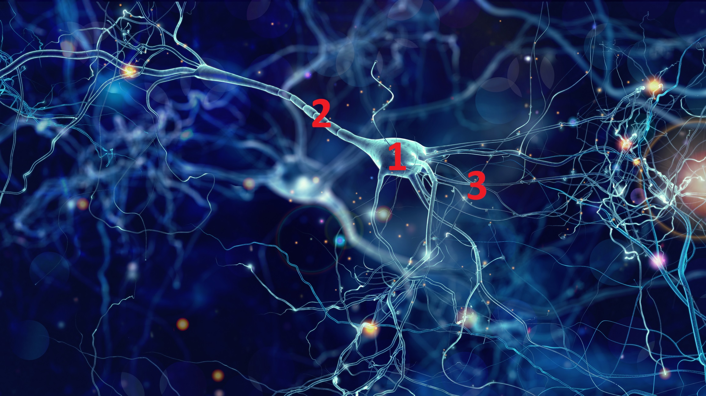
Neurons are made up of three parts.
- A cell body, also called soma (from the Greek somâ meaning body): It is the “thinking head” of the neuron containing a nucleus and an “apparatus” (composed for example of mitochondria which provide the cell with energy) necessary to its proper functioning. It also provides most of the nutritional needs for keeping the neuron alive. The disruption of this apparatus leads to the death of the neuron; it is at the origin of neurological diseases such as Alzheimer’s disease or Parkinson’s disease which are qualified as neurodegenerative diseases.
- An axon (from the Greek axôn meaning axis): it is a single arm which starts from the cell body and ramifies at its end. it is also called nerve fiber. Its length varies from a few millimeters, as in the case of small neurons in the brain, up to one meter in the case of axons that start from the spinal cord to innervate the feet.
- Dendrites (from the Greek meaning dendron shaft): fine and short extensions forming a tree around the cell body. Most of the signals transmitted by neurons enter through dendrites, while other signals enter the cell body. Signals from neurons and projecting onto other neurons go through contact points called synapses. There is therefore no direct contact between neurons.
Thus, the neuron can receive information, store it and transmit it to other neurons.
The functioning of a neuron depends on its ability to communicate with its peers. This communication takes place using points of contact called synapses (from the Greek sun and aptein meaning respectively with and to join). A neuron has several thousand synapses located mainly between the axon of one cell and the dendrites of another cell.
During normal aging, synapses change morphology but remain relatively intact, while their number significantly decreases in the brains of people with Alzheimer’s disease.
The different types
There are different types of neurons depending on the size and shape of the soma, the density of the dendrites, as well as the length and branching of the axon.
Interneurons
Interneurons are found throughout the human body. Interneurons allow communication between sensory or motor neurons and the central nervous system.
Unlike the peripheral nervous system, the central nervous system – which includes the brain and the spinal cord – contains many interneurons. In the neocortex, which makes up about 80% of the human brain, 20% to 30% of neurons are interneurons.
Motor neurons
They have a cell body located in the brainstem, motor cortex, or spinal cord of the body. Its axon, or fiber, protrudes either on the spinal cord or outside the spinal cord in order to directly or even indirectly control the effector organs; in other words, the glands and the muscles.
A single motor neuron can innervate many muscle fibers.
Sensory neurons
They are nerve cells located in the nervous system and responsible for converting external stimuli from the body’s environment into internal electrical impulses.
This process is among the functions that include muscle contractions and even involuntary behaviors such as pain avoidance. These reflex circuits are usually found in the spinal cord in humans.
Also known as afferent neurons, sensory neurons convert a particular type of stimulus into action potentials through their receptors.
Information travels from the sensory nerve to the brain via the spinal cord. Stimuli can come from outside the body, including sound and light; or inside the body, including blood pressure or the sense of body position. Different types of sensory neurons have different receptors that respond to different types of stimuli.
The different types and functions include:
- Taste : found in the taste receptors of the taste
buds. - Odor : olfactory receptor neurons that are activated by odor molecules present in the air.
- Vision : use of photoreceptor cells that convert light into electrical signals.
- Auditory : responsible for converting pressure waves from air molecules or sound into signals that can then be interpreted by the brain.
- Temperature : sensory receptors that react to varying temperatures.
- Mechanoreceptors : These are sensory receptors that respond to mechanical forces, including distortion or pressure.
Do neurons have the same morphology?
No. Neurons, basic elements of the brain, have a very variable morphology. They are distinguished, among other things, by:
- the shape of their cell body;
- the organization and morphology of extensions (i.e. dendrites and their axons) emerging from the cell body.
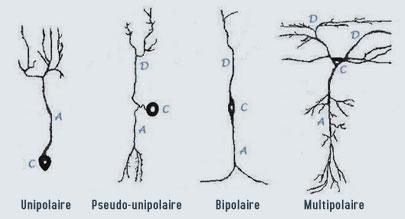
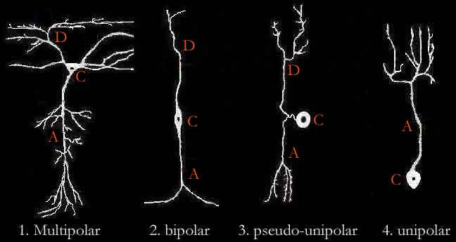
Here are schematically the four types of neurons found in the central nervous system (together making up the brain and spinal cord):
- Unipolar neurons are found mostly in insects, with the cell body located outside the brain. These neurons have only one long extension (the axon) extending from the cell body (C).
- Pseudo-unipolar neurons are sensory neurons located in the spinal ganglion. They pick up information from sensory receptors (e.g. pain receptors) through its dendrites and communicate it via its axon to the dorsal root of the spinal nerve to the spinal cord. The single dendrite (D) and the axon (A) are joined close to the cell body before they separate.
- Bipolar neurons are rather rare sensory neurons specialized in the transmission of the senses (smell, sight, taste, hearing) and vestibular functions (balance, detection of movement). They are found in particular in the retina and the olfactory mucosa. The axon and the single dendrite are opposed to the cell body.
- The multipolar neuron (4) is distinguished as well by the shape of its cellular body …… as by the shape of its axon or its dendrites. The multipolar neuron has a single axon and imposing dendrites (we speak of « dendritic tree »). Multipolar neurons constitute the majority of neurons in the brain.
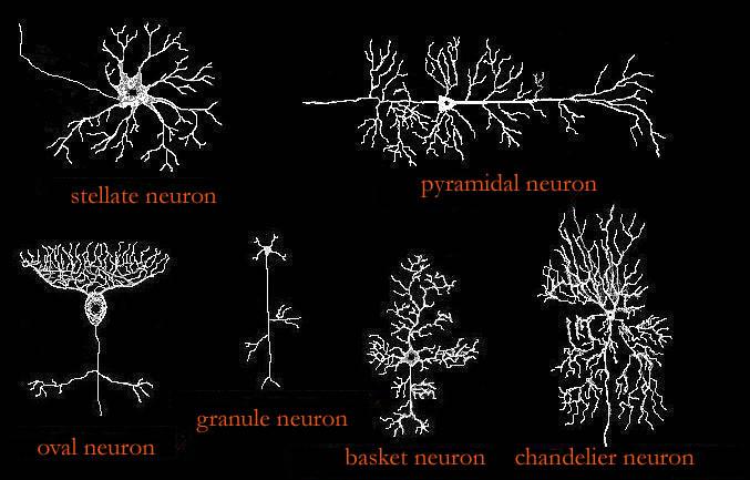
Different types of multipolar neurons
- The axon of a neuron can be long (from a few centimeters to a few tens of centimeters): it is the type I Golgi neuron. It may, for example, be a pyramidal cell, the cell body of which, located in the cerebral cortex, may extend to the spinal cord.
- The neuron can have a short axon (a few hundredths of a millimeter) that divides rapidly to give a complex arborization. This type of neuron remains within the limits of the structure in which it is found: it is the Golgi type II neuron. It can be, for example, a neural cell in a basket or in a star.
- For some neurons, the axon and its collaterals present swellings called “spider veins” along their path. For other neurons, dendrites have irregular contours (called « dendritic spines »).
Multipolar neurons are, for example, motor neurons responsible for motor control from the cortex to the spinal cord or from the marrow to the muscles, but also interneurons which conduct impulses inside the central nervous system. Multipolar neurons make up the majority of neurons in the brain.
In summary, there are different types of neurons that come in very different forms. This polymorphism has an impact on the ability of the neuron to receive and process the information it receives from another neuron.
The multipolar neuron’s (1) morphology is diverse and is distinguishable not only from the shape of the cell body, but also from the shape of the axon or dendrites.