La neuroinflammation

White matter

The limbic system

The hippocampus and its role in the brain

Leukopathy

Basal ganglia

What are the cranial nerves?

Xanax: how long does its effect last?

Nouveau jeu aviator au casino

Quels sont les effets secondaires du vaccin contre le COVID ?

Author Archives: Stéphane Bastianetto

  1. La neuroinflammation

    Commentaires fermés sur La neuroinflammation

    La neuroinflammation est une réaction inflammatoire qui se déroule dans le cerveau et au cours de laquelle s’expriment des molécules appelées cytokines.

    Les tissus réagissent en réponse à une infection ou à la présence d’un corps étranger : cette réaction inflammatoire se caractérise par des rougeurs ou une lésion fonctionnelle.

    Les acteurs participant à ces phénomènes sont les globules blancs (appelés leucocytes) produits par la moelle osseuse et présents dans le sang.

    Le nombre des leukocytes augmente lors de l’inflammation.

    Il existe trois grandes classes de globules blancs : les lymphocytes, les granulocytes et les monocytes.

    • Les lymphocytes participent à la réaction immunitaire en produisant des anticorps qui vont aider à détruire l’agent pathogène (par ex. une bactérie).
    • Les granulocytes qui sont des globules blancs qualifiés de « non spécifiques » à un antigène.
    • Les monocytes sont des cellules du sang qui phagocytent (du grec phagos, manger; la phagocytose a été mise en évidence au début du XXème siècle par le biologiste russe Elie Metchnikoff), c’est-à-dire qu’ils avalent et digèrent les débris cellulaires et les particules vivantes (par ex. des microbes).

    Il existe un autre type de cellules « dévoreuses » ayant les mêmes propriétés que les monocytes: ce sont les macrophages (du grec makros, grand). Alors que les monocytes se situent spécifiquement dans le sang, les macrophages sont localisés dans les tissus.

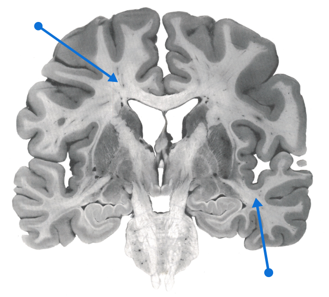
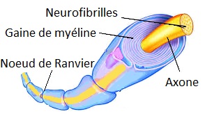
    Il existe ainsi des macrophages dans le cerveau qui portent le nom de cellules microgliales: elles forment avec les astrocytes et les oligodendrocytes les cellules gliales (lire l’article sur le (article sur le fonctionnement du cerveau).

    Le cerveau peut-il être la cible d’une inflammation ?

    On croyait jusqu’à récemment que le cerveau bénéficiait -contrairement aux autres organes- d’un privilège immunitaire et qu’il échappait largement à la surveillance immunitaire. Il n’en est rien.

    La cellule microgliale joue à ce titre un rôle évidemment central puisque c’est la cellule qui phagocyte les éléments étrangers. Mais elle peut également se retourner contre les cellules voisines, c’est-à-dire les neurones, notamment lors de troubles neurologiques. C’est en quelque sorte Dr. Jekyll et Mr. Hyde.

    Ainsi la maladie d’Alzheimer s’accompagne d’une activation des cellules microgliales. Cette réaction provoque une réaction inflammatoire appelée neuroinflammation, au cours de laquelle s’exprime une bonne centaine de molécules différentes et que l’on appelle cytokines.

    Les cytokines

    La caractéristique d’une réaction immunitaire et d’une neuroinflammation est la production de cytokines (du grec kutos, cellule, et kinéo, stimuler), messagers chimiques permettant aux cellules de communiquer entre elles.

    Elles sont donc libérées par les cellules gliales lorsqu’un agent infectieux ou toxique attaque l’organisme.

    Les cytokines vont ainsi induire, contrôler ou inhiber l’intensité et la durée de la réponse immunitaire. Lorsque les cytokines sont sécrétées par les cellules, elles agissent:

    • en circulant dans le sang (c’est le mode endocrine, du grec endon, au-dedans; ekkrinein, excréter),
    • en agissant sur les cellules qui les sécrètent elles-mêmes (c’est le mode autocrine, du grec auto, en soi) ou
    • en agissant sur les cellules voisines (c’est la mode paracrine, du grec para, à côté de).

    Elles sont impliquées dans un grand nombre de fonctions, en particulier dans la résistance aux agents infectieux ou toxiques. Elles englobent donc plusieurs domaines d’application : cancérologie, hématologie, immunologie, infectiologie et neurologie. Il existe une quarantaine de cytokines identifiées à ce jour, regroupées en familles. Celles jouant un rôle dans les troubles cérébraux sont :

    – Les interleukines (IL) telles que l’interleukine 1 (IL-1).

    – Le transforming growth factor-ß.

    – Le facteur de nécrose tumorale (ou TNF-alpha pour tumor necrosis factor-alpha).

    De nombreuses études ont montré que de nombreuses agressions au cerveau venant de traumatismes crâniens, d’accidents vasculaires cérébraux, d’infections ou de certaines maladies mentales (schizophrénie, maladie d’Alzheimer) sont associées à de fortes concentrations de cytokines (par exemple IL-1 ou TNF-alpha).

    Ce phénomène de surproduction de cytokines dans le cerveau s’appelle la neuroinflamation.

    La neuroinflammation est impliquée dans la maladie d’Alzheimer

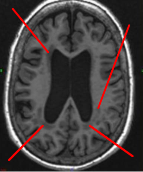
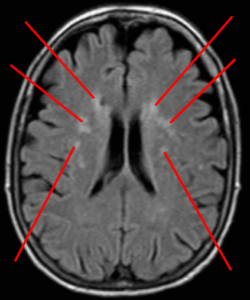
    La neuroinflammation se caractérise donc par une libération accrue de certaines cytokines dans le cerveau de patients atteints de la maladie d’Alzheimer (MA). Une de ces cytokines serait particulièrement impliquée dans la MA: la TNF-alpha. En effet :

    – Les niveaux de TNF-alpha sont 25 fois plus élevés dans le liquide céphalorachidien (liquide dans lequel baigne le cerveau) des patients MA que chez les personnes saines; de plus les patients souffrant de déclin cognitif léger et ayant des niveaux élevés de TNF-alpha ont un risque accru de développer une MA.

    – Des polymorphismes génétiques* associés à une augmentation de la production de TNF-alpha ont été observés chez certaines populations ayant un risque accru de développer une MA.

    * Polymorphismes génétiques : un gène a deux copies appelées allèles. Ces allèles peuvent exister sous différentes formes dans une population : c’est le polymorphisme. Cette différence d’expression n’engendre pas une maladie mais peut augmenter le risque de la développer.

    – Selon une étude épidémiologique, une production de TNF-alpha par des cellules sanguines (macrophages, mastocytes) est associée à une augmentation du risque de développer une MA.

    – Les études réalisées chez l’animal renforcent l’hypothèse d’un rôle de la TNF-alpha dans le dysfonctionnement des synapses (zones de contact entre les neurones) lors du vieillissement pathologique; en particulier lorsque les synapses sont exposées à l’amyloïde, protéine jouant un rôle inhibiteur dans la mémoire et dans la mort neuronale dans la MA.

    Il a également été montré que le TNF-alpha et l’amyloïde rentrent dans un cercle vicieux dans lequel l’amyloide stimule les cellules microgliales, ce qui excite neurones et cellules gliales, qui fabriquent d`avantage de l’amyloïde, et ainsi de suite.

    TNF-alpha et la maladie d’Alzheimer

    L’excès de TNF-alpha produite par les macrophages va provoquer une réaction inflammatoire excessive qui va endommager les articulations et provoquer des rhumatismes inflammatoires telle que la polyarthrite rhumatoïde. Des molécules capables de bloquer l’action des TNF-alpha ont déjà été développées dans le traitement de cette maladie. Ces molécules sont : étanercept, infliximab et adalimumab. En se basant sur ces résultants, la communauté médicale s’est proposé de tester l’une de ces molécules chez des patients Alzheimer.

  2. White matter

    1 Comment

    White matter is the part of the tissue of the central nervous system (including the brain and spinal cord) that mainly contains extensions of neurons.

    These extensions called axons are surrounded by a myelin sheath which is the origin of this whitish structure, as well as glial cells which nourish and protect the nerve cells.

    The myelin is a fat which protects the fibers and gives color to the white matter.

    It also ensures rapid propagation of information in the nervous system in the form of an electrical signal. These lesions are characterized by damage to the tissues located in the deepest part of our brain, due to health problems associated with aging. This tissue contains millions of axons, which connect other parts of the brain and spinal cord. 

    Frontal section of a human brain with white matter (blue arrows)

    White matter lesions

    Lesions from this substance are common in the elderly, especially in those with a history of hypertension and cerebral deficiencies.

    Many diseases, injuries, and toxins can cause white matter changes:

    • Long-term high blood pressure
    • Inflammation of the blood vessels
    • Tobacco consumption

    Controlling hypertension well

    The results showed that individuals with white matter lesions more often had a history of hypertension (+ 270%), compared to those without lesions. They also had a much larger number of brain deficiencies (+ 448%).

    Cognitively, patients with these types of lesions performed poorer on cognitive tests, suggesting that this category of people has an even greater risk of developing Alzheimer’s disease.

    « Our results report a correlation between white matter lesions and impaired cognitive functions, suggesting that these lesions worsen this deficit in individuals with mild cognitive impairment. »

    Consequently, it is advisable to control the cardiovascular risk factors at the origin of these lesions, in particular hypertension, and to a lesser extent diabetes, hypercholesterolemia and atheroma.

    It remains to be determined whether these lesions increase the conversion rate between mild cognitive impairment and dementia.

    Source: Duron E. et al. Relations between cognitive disorders and lesions of the cerebral white matter. Archives of diseases of the heart and vessels. 100 (8), 2007.

    Symptoms

    • Learning and memory disabilities: difficulty learning or remembering new things
    • Difficulty solving problems
    • Incontinence
    • Depression
    • Walking disorders
    • Balance disorders with risk of falls

    People with damage to white matter – the part of the brain made up of the axons of neurons – have an increased risk of stroke and dementia such as vascular dementia or Alzheimer’s disease. It is possible to prevent and slow down this damage.

    (1) Gray substance; (2) White substance

    White matter lesions increase risk of stroke

    A meta-analysis comprising twenty-seven studies that compared healthy older people to those with cognitive impairment caused by white matter damage.

    White matter lesions affect small blood vessels deep in the brain, unlike Alzheimer’s disease which narrows the hippocampus, causing progressive loss of memory.

    The disease hardens the small arteries and gradually limits the arrival of nutrients in the white matter, leading to a loss of several cognitive functions: planning, organization, problem solving, attention, working memory, immediate and delayed memory.

    These conditions increase a person’s risk of having a stroke and even of developing dementia such as vascular dementia or Alzheimer’s disease.

    “Our results support the idea that white matter lesions gradually alter the brain and cognitive functions, without modifying activities of daily living  », declared the author of a study published in 2014.

    White matter damage increases risk of Alzheimer’s

    In 2004, a Dutch study involving more than a thousand subjects followed for 5 years already indicated that white matter (WM) damage quadrupled the risk of dementia, including Alzheimer’s type dementia ( Prins et al., Arch Neurol., 61: 1531- 4 ).

    It is estimated that 5 percent of Canadian adults have a vascular cognitive impairment, possibly related to hypertension , high cholesterol , poorly controlled diabetes, poor dietary hygiene, inactivity and smoking.

    WM damage is anatomically characterized by arteriosclerosis, myelin damage and activation of glial cells (gliosis). They are caused by an ischemic attack which affects the small arteries.

    The good news is that WM damage can be reduced by adopting healthy lifestyle habits that reduce the risk of heart attack and stroke.

    Source:  The neuropsychological profile of vascular cognitive impairment not demented: A meta-analysis. Journal of Neuropsychology, février 2014.

    In another study, American researchers reported white matter and gray matter damage in people aged 40. This damage is subsequently associated with a decline in cognitive functions, or even with Alzheimer’s disease.

    The study’s author, Dr. Pauline Maillard, explained that the study shows for the first time that hardening of the arteries harms the brain and that the problem manifests itself in midlife, much earlier than did not anticipate it.

    Worsening of cognitive impairment

    White matter lesions, common in older people with hypertension, may be responsible for mild cognitive impairment.

    Another study determined whether the presence of WM damage was associated with a more marked decline in cognitive performance in individuals with mild cognitive impairment, a sometimes transient state between normal cognitive functioning and dementia. Almost 15% of people with mild cognitive impairment (MCI) develop dementia per year.

    136 patients – diagnosed with mild cognitive impairment – underwent a battery of neuropsychological tests * and a neurological examination (CT scan) to determine the presence of lesions.

    * Mini-examination of mental state and cognitive efficiency profile

    These patients were on average 75 years old, of whom more than half had hypertension (54%) and a third had white matter lesions. Hypertension was characterized by a pressure greater than 140/90 mmHg.

    How are they diagnosed?

    A magnetic resonance imaging ( MRI ) test can show any damage. White matter changes appear to be very bright (referred to as a « hyper-intense » signal) during an MRI scan.

  3. The limbic system

    Leave a Comment

    The limbic system is one of the oldest parts of the brain. It is present in humans, but also in reptiles and fish.

    A term introduced by Paul MacLean in 1952, the limbic system was long considered the seat of emotions (aggressiveness, fear, pleasure, anger), representing the dialogue between the brain and the body. 

    Functions of the limbic system

    The limbic system is not only involved in emotions but also in:

    • memory learning,
    • olfaction,
    • the control of the endocrine system which participates in the release of hormones,
    • eating behaviors and appetite,
    • the autonomic nervous system which controls respiratory, digestive and cardiovascular functions.

    Anatomy of the limbic system

    The limbic system is made up of several nuclei located under the cortex (they are said to be subcortical structures) and near the thalamus:

    • The hippocampus (from the ancient Greek hippocampos, meaning « bent horse »): role in learning and storing information in long-term memory.
    • The amygdala (from the Latin amygdala which means « almond »): role in aggression, anger, fear, anxiety and emotional memory. The German physiologist Burdach (1776-1847) is the origin of the term  amygdala. When electrically stimulated, animals become aggressive. And if the amygdala is removed, the animals become very tame and no longer respond to things that would have caused rabies before.
    • The fornix .
    • The limbic cortex (cingulate gyrus, cingulum, insula and parahippocampal gyrus): role in the conscious control of behavior.
    • The septum. One of the first functional roles to be associated with the septum was involvement in the reward (or reinforcement) circuit. The nuclei of the septum have been implicated in a number of other roles such as social behavior and fear expression, and abnormalities in septal functioning have been linked to a variety of illnesses ranging from depression to schizophrenia.
    • The hypothalamus. It is a vital part of the limbic system which is responsible for the production of multiple chemical messengers, called hormones. These hormones control the body’s water levels, sleep cycles, body temperature, and food intake. The hypothalamus is located below the thalamus.
    • The mammillary bodies.
    • The cingulate gyrus. It serves as a channel for transmitting messages between the inner and outer parts of the limbic system.
    • Anterior nucleus of the thalamus.
    • The epiphysis

    The limbic system is involved in emotions and memory

    The limbic system is involved in the feeling of fear which can be reproduced by stimulating through the hypothalamus and amygdala. Conversely, by destroying the tonsils, the fear and the reaction on the body disappears. For example, when a walker is surprised in the forest by a snake, his tonsil is stimulated, which causes an increase in heart rate and stress.

    The limbic system (especially the tonsils) is also the source of anger , as is the phenomenon of addiction (drug use) and pleasure (eg consumption of sugars).

    Finally, the tonsils and the hippocampus are involved in the formation, maintenance and extinction of phobic memory (eg fear of certain animals).

    The main components

    The hippocampus  receives neurons from the limbic cortex, one of the components of which is the parahippocampal gyrus. The parahippocampal gyrus includes the entorhinal cortex, one of the first areas of the brain affected in Alzheimer’s disease. The limbic cortex in turn receives afferents * from other associative cortices (parieto-temporo-occipital and prefrontal).

    Afferences: Scientific jargon meaning that the limbic cortex receives neurons from other cortices. We can also say that the associative cortices send efferences towards the limbic cortex, or that they project towards the limbic cortex.

    Neurons from the hippocampus project via the fornix into the hypothalamus and the septal nucleus. They also project into the entorhinal cortex which in turn projects into the different cortices (prefrontal, orbital, parahippocampal, cingulate, insular). The mammillary nucleus projects onto the mamillothalamic tract which in turn projects onto the cingulate gyrus. A bidirectional circuit is thus formed: it is the circuit of Papez. It plays a central role in memory .

    The amygdala is a set of subcortical nuclei (basolateral, central and corticomedial nuclei) located in the medial temporal lobe. The basolateral nuclei receive projections from the sensory and associative cortical areas of the temporal lobe. They in turn project to the limbic cortex, the prefrontal cortex and the hippocampal formation. The basolateral nuclei project to the thalamus, which projects to the prefrontal cortex. Basolateral nuclei also project to Meynert’s basal nucleus, a nucleus made up of cholinergic neurons that send neuronal projections to the cortex (these neurons are damaged in Alzheimer’s disease). The basolateral nuclei of the amygdala are thought to be involved in the emotional component of sensory stimuli, as well as the memorization of emotional stimuli.

    Located below the thalamus, the hypothalamus  sends out neurons that control:

    • the secretion of certain hormones secreted by the pituitary gland (or pituitary gland),
    • the autonomic nervous system (regulation of temperature, the circadian cycle, heart rate, sweating), and
    • certain behaviors (sexual, food, defense, stress).

    The basal ganglia

    The limbic system is associated with the basal ganglia (or basal ganglia ), a region of the brain comprising a collection of subcortical nuclei located approximately in the center of the human brain. These nuclei are the striatum (grouping together the caudate nucleus and the putamen), the internal and external globus pallidus, the subthalamic nucleus and the substantia nigra.

    Connected with the cerebral cortex and the thalamus, they play a fundamental role in voluntary motor skills but also in learning, memory and emotions.

    The role of the basal ganglia in emotions is explained by the fact that the ventral part of the striatum receives neuronal projections from regions of the limbic system (amygdala, hippocampal formation and limbic associative cortex).

    Neurons from the globus pallidus project into the thalamus, which in turn projects onto the prefrontal cortex.

    The limbic system includes cortical and subcortical brain areas involved:

    • in declarative memory and learning (hippocampus and parahippocampal gyrus) and,
    • emotions and behavior (amygdala).

    Note that the amygdala is also involved in emotional memory.

    Let’s remember that :

    • the diencephalon is made up of the thalamus (from the Greek meaning « chamber »), the hypothalamus and the subthalamus.
    • The telencephalon is made up of the hippocampus (composed of the hippocampus, subiculum and dentate gyrus), amygdala, striatum (caudate nucleus and putamen) and limbic cortex.
    Frontal section of a human brain. The regions of the limbic system (limbic cortex, hippocampus) coexist with those of the basal ganglia (caudate nucleus, putamen, substantia nigra.

    Legend. Limbic cortex (1); frontal lobe (2); parietal lobe (3); island lobe (4); temporal lobe (5); temporal gyrus (6); hippocampus (7); parahippocampal gyrus (8); cranial nerves (9); substantia nigra (10); red core (11); putamen (12); caudate nucleus (13).

  4. The hippocampus and its role in the brain

    Leave a Comment

    The hippocampus is a structure of the brain that plays a fundamental role in learning and memory. It is damaged in Alzheimer’s disease.

    Knowing about the hippocampus has helped researchers understand how memory works.

    Location

    The hippocampus is a structure of the brain that is part of the temporal lobe of the cerebral cortex.

    The name comes from the Greek words hippo, meaning horse, and kampo, meaning monster, because its shape resembles that of a sea horse. It has a C shape.

    The hippocampus is a brain structure located in the temporal lobe. It plays a major role in learning and memory.

    The hippocampus is part of the limbic system. It is found under the cortex.

    The limbic system is considered a « primitive brain », located deep in the brain. It is involved in hunger, motivation, libido, mood, pain, pleasure, appetite and memory, etc.

    The hippocampus is the part of the brain that is one of the most widely studied. Its atrophy has clinical consequences.

    It is the earliest and most severely affected structure in several neurological disorders such as Alzheimer’s disease or epilepsy.

    In adults, the volume of the hippocampus on each side of the brain is approximately 3-3.5 cm 3 while the volume of the cerebral cortex is approximately 320-420 cm 3.

    Thus, the hippocampus is 100 times smaller than the cerebral cortex.

    Papez (1930) proposed that the emotional reaction is organized in the hippocampus and is expressed in the cingulate gyrus. He is also involved in recalling past experience and how to imagine the future.

    But its best-known role is that in learning and short-term memory.

    Anatomy

    The hippocampus contains two parts: the Cornu ammonis or horn of Ammon (with its CA1, CA2, CA3 and CA4 regions) and the dentate gyrus. These two parts are separated by the sulcus. Finally there is the entorhinal region (EC). 

    The whole is called hippocampal formation.

    The hippocampus is divided into a head, a body and a tail; the head being an enlarged part while the tail is a thin part.

    Just in front of CA1 is the subiculum that connects the hippocampus to the entorhinal cortex.

    The hippocampus is supplied with blood by the posterior cerebral artery, which has three branches: anterior, middle and posterior. The veins of the hippocampus go through the basal vein.

    Hippocampus and memory

    The hippocampus can process and recover two types of memory, episodic memory and spatial memory.

    The episodic memory is related to facts and events. 

    Spatial memory involves paths or routes. For example, when a taxi driver learns a route through a city, he uses spatial memory. 

    The hippocampus is also where short-term memories are turned into long-term memories. These are then stored elsewhere in the brain.

    Research has shown that neurons continue to develop throughout adulthood. The hippocampus is one of the rare places in the brain to generate new nerve cells: it is neurogenesis.

    The HM case

    The role of the hippocampus in memory was particularly revealed in the case of Henry Gustav Molaison (called HM). Partial anterograde and retrograde amnesia developed in this patient following the removal of his hippocampus due to refractory epilepsy. HM was unable to form new episodic memories after this surgery. In other words, he was no longer learning new things and not remembering what he had learned before the operation. In medical science, HM is perhaps the most studied patient. 

    Subsequent studies have shown that damage to the hippocampus causes anterograde amnesia and often retrograde amnesia as well. Implicit memory is spared due to damage to the hippocampus.

    The hippocampus is now known not only to be important in learning and memory, but also in:

    • Orientation in space
    • Emotional behavior.

    The regulation of the functions of the hypothalamus and consequently the release of cortisol, the stress hormone.

    Hippocampus and memory loss

    Transient global amnesia is a specific form of memory loss that develops suddenly, apparently on its own, and then goes away fairly quickly.

    Most people with transient global amnesia eventually regain their memories, but it’s unclear why the problem occurs and why it resolves. Damage to the hippocampus may be involved.

    Damage to the hippocampus can make it difficult to remember how to get from one place to another. The person may be able to draw a map of the neighborhood they lived in as a child, but it may be difficult for them to go to a store in a new neighborhood.

    It has also been linked to diseases such as schizophrenia and post-traumatic stress disorder .

    Lesions of the hippocampus associated with diseases of the brain

    The hippocampus is a sensitive part of the brain. A range of illnesses can negatively affect it, including long-term exposure to high levels of stress.

    Several diseases and factors are known to interfere with the ability of the hippocampus to function normally.

    Alzheimer’s disease

    The atrophy of the hippocampus region of the brain is one of the features most consistent with Alzheimer’s disease. It is the most severely affected region of the brain. 

    One hypothesis has proposed that early lesions of the hippocampus cause « dissociation » between the hippocampus and the cerebral cortex, leading to failure to record information from the hippocampus. Hippocampal atrophy is used as both a diagnostic and prognostic marker in clinical trials for Alzheimer’s disease. It has been observed that patients with mild cognitive impairment have 10-15% loss of hippocampal volume while in those with early-onset Alzheimer’s disease this loss is approximately 15-30%. In those with moderate Alzheimer’s disease, it can reach 50%.

    Depression

    It is increasingly recognized that prolonged depression can lead to loss of hippocampal volume. In addition, the duration of depression was correlated with the severity of hippocampal atrophy. Evidence suggests that the atrophy so produced may be permanent and persist for a long time even if there is remission. In people with depression, the hippocampus can shrink by up to 20%, according to some researchers. It has been speculated that this could be the result of prolonged stress generated by depression. Suppression of neurogenesis (production of new neurons) in the hippocampus could be the cause.

    Schizophrenia

    Reduction in hippocampal volume is one of the findings observed by MRI in schizophrenic patients. It is less marked than that observed in Alzheimer’s disease. 

    Epilepsy

    Up to 50% to 75% of patients with epilepsy may have atrophy of the hippocampus during postmortem analysis. However, it is not clear whether epilepsy occurs as a result of hippocampal sclerosis or repeated seizures damaging the hippocampus. This means that it is not yet clear whether damage to the hippocampus is a cause or a consequence of recurrent seizures.

    The hippocampus is believed to have an inhibitory effect on the seizure threshold (i.e., it maintains the threshold high). Once it is damaged, the seizures become more uncontrollable.

    Cushing’s disease

    Cushing’s disease has a number of symptoms related to high levels of cortisol, a hormone produced when people are under stress. 

    A loss of hippocampal volume has been observed in Cushing’s disease. There is evidence to suggest that if Cushing’s disease is treated, the atrophy of the hippocampus may be reversible.

    Ongoing research

    In 2016, scientists published a review of studies on the effects of exercise on cognitive decline and aging of the brain.

    The results suggest that exercise may enhance the ability of this structure to generate new nerve cells (neurogenesis). This would preserve and potentially improve memory. This hypothesis remains to be confirmed.

    Conclusions

    The hippocampus, located in the temporal lobe, plays a central role in memory. It is a vulnerable and plastic structure, which can change. Its atrophy is one of the markers of cognitive decline and the diagnosis of Alzheimer’s disease.

  5. Leukopathy

    Leave a Comment

    Leukopathy is a neurological disorder characterized more precisely by a lesion of the white matter whatever the cause of the lesion.

    Leukopathy comes from the Greek leuco = white and from pathos = disease. 

    Leukoencephalopathy is also a medical term used by the physicians.

    Most of the time, it is accompanied by blood circulation problems in the brain. The latter is then poorly irrigated by the arterioles and  capillaries. We speak of leukopathy (or vascular leukoencephalopathy).


    What is white matter?

    White matter is part of the nervous tissue made up of axons. Axons are extensions of neurons  that allow information to pass between two neurons by propagating nerve impulses .

    They are surrounded by a myelin sheath, a lipid layer (made of cholesterol, phospholipids and glycolipids.

    White matter is therefore involved in the transmission of information in the central nervous system and the brain in particular.

    The myelin sheath is therefore damaged in leukopathy.

    Axon surrounded by a myelin sheath. This sheath is damaged in vascular leukopathy


    Classification of different types of leukopathy

    A distinction is made between acquired leukopathies and those which are hereditary.

    Hereditary leukoencephalopathies having a genetic cause are then classified according to the gene which is involved (this gene mutates and this mutation is at the origin of the lesion of the white matter).

    As for acquired leukopathies, they are classified into different groups according to their underlying causes:

    • inflammatory and non-infectious.
    • inflammatory and infectious.
    • hypoxic and ischemic. (example Binswanger’s disease)
    • traumatic.
    • toxic and metabolic.

    Examples of diseases that lead to leukopathy

    There are several diseases that lead to white matter damage :

    Multiple sclerosis

    The multiple sclerosis is a neurological disease characterized by inflammation of neurons (called neuroinflammation).

    Progressive multifocal leukoencephalopathy

    Progressive multifocal leukoencephalopathy (PML) is a rare, often fatal neurological disorder characterized by nerve cell destruction caused by the JC virus. It has been associated mainly with hematological malignancies and organ transplant recipients, but the number of cases has increased significantly due to the use of immunosuppressive drugs. Common symptoms include cognitive dysfunction, hemianopsia, aphasia, motor or sensory dysfunction, seizures and ataxia. PML starts as an asymptomatic persistent infection in the kidney and may spread to other organs through contact with an infected individual or contaminated food or water. The syndrome was particularly common among people with HIV/AIDS in the 1980s, but its occurrence has increased significantly since then due to the wider use of immune-modulating drugs such as natalizumab and efalizumab.

    Leukoaraiosis

    Leukoaraiosis is also accompanied by neuroinflammation.

    Alzheimer’s disease

    The Alzheimer’s disease is the most important form of dementia and is accompanied by abnormalities of the white matter.

    Toxic leukoencephalopathy

    It is a rare condition characterized by progressive damage to the white matter caused by drug addiction, environmental toxins, or chemotherapy drugs. The severity of toxic leukoencephalopathy also varies depending on the patient, the duration of exposure and the concentration of the toxic agent. The disease may be reversible in many cases when the toxic agent is removed.

    Reversible posterior leukoencephalopathy syndrome

    Posterior Reversible Encephalopathy Syndrome (PRES) is a syndrome characterized by headache, confusion, seizures and loss of vision. It can have several causes, including malignant hypertension and certain medical treatments. On magnetic resonance imaging (MRI) of the brain, there are areas of edema (swelling). Symptoms tend to go away after a while, although sometimes visual changes remain.

    Hypertensive leukoencephalopathy

    Hypertensive leukoencephalopathy refers to a degeneration of the white matter of the brain following a sudden increase in blood pressure. People may experience a sudden increase in blood pressure, an acute confusional state, headache, vomiting, and seizures. Retinal hemorrhages may be present during the examination. Hypertensive leukoencephalopathy can lead to cardiac ischemia.

    Along with these acquired leukoencephalopathies, there are those which originate from a genetic mutation:

    Megalencephalic leukoencephalopathy with subcortical cysts. This form of leukopathy has a genetic cause. It is characterized by subcortical cysts (small cavities). It belongs to a group of diseases called leukodystrophies. (also called Van der Knaap disease).

    Diffuse hereditary axonal spheroid leukoencephalopathy 

    Leukoencephalopathy with neuroaxonal spheroids is a special form of leukoencephalopathy. Spheroids are discontinuous axonal swellings characterized by the absence of myelin sheaths. They cause progressive cognitive and motor regression. It is a disease of genetic origin, following an autosomal dominant profile . It usually presents in childhood, but it can also appear in adulthood, in which case it can have neurological features similar to those of multiple sclerosis. 

    Hereditary vascular leukoencephalopathies

    They include CADASIL disease (NOTCH3 gene mutation), CARASIL disease (HTRA1 gene mutation) and cerebral amyloid angiopathy. In the case of CADASIL, there is a family history of cerebral stroke.

    Childhood Ataxia with diffuse Central nervous system Hypomyelination

    This syndrome was initially described in children. The adult forms
    are however more and more recognized. The neurological symptoms are very variable (cerebellar ataxia, psychiatric and dementia symptoms). It is an autosomal recessive neurological disease, caused by mutation of EIF genes. This disease belongs to the leukodystrophies family.

    How to screen for vascular leukopathy?

    Brain imaging exams (MRI, scanner) make it possible to detect leukopathies. These leukopathies result in lacunae (small cavities that reflect damage to the central nervous system), small bleeding and damage to the outer capsules of the white matter.

    Leukopathies generally affect people with vascular risk factors (with a risk of vascular dementia), with cardiovascular problems or suffering from atherosclerosis. These are, for example, people with diabetes, hypertension and high cholesterol.

    It is possible that individuals who are free of any of the diseases described below and under the age of 50 may develop leukopathy. In this case, the diagnosis will be based on a genetic disease such as leukodystrophy, also called genetic leukoencephalopathy.

    Leukoencephalopathies of genetic origin include hereditary diseases characterized by a disease of the white matter:

    • vascular leukoencephalopathies such as CADASIL disease and familial cerebral amyloidosis.
    • Ataxia with Central nervous system hypomyelanisation.
    • Fragile X syndrome with early gait disturbances.
    White matter damage appearing as black spots on a horizontal section of a patient’s brain. These attacks are visualized by the MRI technique
    Moderate white matter lesions visualized on a horizontal section of a brain. This time, the MRI technique makes it possible to detect these lesions in the form of white spots.

    The consequences of leukopathy

    Leukopathy increases the risk of stroke, cognitive impairment (memory, executive functions), movement coordination disorders or seizures.

  6. Basal ganglia

    Leave a Comment

    The  basal ganglia are areas of the brain located below the cortex, deep in both hemispheres of the brain . These nuclei are involved in the control of voluntary motor movements, learning and decision-making on the performance of motor activities.

    Diseases that affect this region include Parkinson’s disease  and Huntington’s disease.

    They are surrounded by white matter from the cerebral hemisphere.

    Divisions of the basal ganglia

    • The striatum (caudate nucleus and putamen)
    • The globus pallidus
    • The amygdala
    • The black substance
    • The subthalamic nuclei
    The basal ganglia (or basal ganglia) and their constituent structures: globus pallidus (1), putamen (2), caudate nucleus (3), subthalamic nucleus (5), nucleus accumbens (6). The structures connected to the basal ganglia are the thalamus (4) and the amygdala (7).
    Frontal section of a brain of a healthy subject representing the striatum including the caudate nucleus and the putamen (1), the thalamus (2) and the frontal lobe (3).

    The basal ganglia receive almost all of their afferent signals from the cortex and send almost all of their efferent signals back to the cortex.

    Functions

    • Play the role of mediator between the motor centers of the neocortex and the motor areas of the brainstem
    • Select the desired and desired motor activity and remove unwanted movements.
    • Ensure an inhibitory role in motor control
    • Inhibit muscle tone
    • Adjust slow and continuous contractions (balance, body position, etc.)
    • Regulate attention and individual cognitive processes
    • Participate in motor planning and learning
    • Help the cerebral cortex to perform subconscious and learned movements

    Neurotransmitters

    • Dopamine producing neurons (dopaminergic neurons) located in the substantia nigra (or substantia nigra) send projections to the caudate nucleus and putamen.
    • GABA-containing neurons (gabaergic neurons) located in the nucleus caudatus and putamen, which send projections to the substantia nigra.
    • Neurons that produce acetylcholine (cholinergic neurons) located in the cerebral cortex, which send projections to the caudate and putamen.
    • Norepinephrine and serotonin neurons send projections to the basal ganglia.
  7. What are the cranial nerves?

    Leave a Comment

    The 12 cranial nerves form a set of nerves that originate in the brain.

    Their functions are sensory, motor or both at the same time:

    • Cranial sensory nerves help a person see, smell, and hear.
    • Cranial motor nerves help control muscle movement in the head and neck.

    Each nerve has a name that reflects its function and a number based on its location in the brain.

    Scientists use Roman numerals from I to XII to identify the cranial nerves in the brain.

    Doctors can identify neurological or psychiatric disorders by assessing the functions of the cranial nerve.

    I Olfactory nerve

    The olfactory nerve transmits information about a person’s smell to the brain.

    When a person inhales scent molecules, olfactory receptors located in the nasal passage send the impulses to the cranium and then travel to the olfactory bulb.

    Olfactory neurons merge with other nerves, which pass through the olfactory tract.

    The olfactory tract then travels to the frontal lobe and other areas of the brain that are involved in memory and detection of different smells.

    II Optic nerve

    The optic nerve transmits information about a person’s vision to the brain.

    When light enters the eye, it hits the retina, which contains rods and cones. These are photoreceptors that translate signals from light into visual information for the brain.

    The cones are located in the central retina and participate in color vision. The rods are located in the peripheral retina and participate in uncolored vision (night vision).

    These photoreceptors carry nerve impulses along nerve cells.

    Most of the optic nerve fibers intersect in a structure called the optic chiasm. Then, they project to the primary visual cortex located in the occipital lobe at the back of the brain. The occipital lobe is where the brain handles visual information.

    III Oculomotor nerve

    The oculomotor nerve is a cranial nerve that helps control muscle movement in the eyes.

    The oculomotor nerve moves most of the muscles that move the eyeball and upper eyelid, called extraocular muscles.

    The oculomotor nerve also contributes to the involuntary functions of the eye:

    The pupillary muscle automatically constricts the pupil to allow less light to pass into the eye when the light is bright. When it is dark, the muscle relaxes to let in more light.

    Ciliary muscles help the lens adapt to short and long distance vision. This happens automatically when a person is looking at near or far objects.

    IV Trochlear nerve

    The trochlear nerve is also involved in eye movement.

    The trochlear nerve, like the oculomotor nerve, originates in the midbrain. It supplies the contralateral superior oblique muscle which allows the eye to point downward and inward.

    V Trigeminal nerve

    The trigeminal nerve is the largest cranial nerve and has motor and sensory functions.

    Its motor functions help a person chew and clench their teeth and contract the muscles of the eardrum.

    It consists of three parts that connect to sensory receptors on the face:

    The ophthalmic part transmits sensation to certain parts of the eyes, including the cornea, the lining of the nose, and the skin of the nose, eyelid and forehead.

    The jawbone transmits sensation to part of the face, the side of the nose, the upper teeth and the lower eyelid.

    The mandibular part transmits sensation to the lower part of the face, tongue, mouth and lower teeth.

    Trigeminal neuralgia can cause severe pain and facial tics.

    VI Nerf Abducens

    The abducens nerve also helps control eye movements.

    It helps the lateral rectus muscle, which is one of the extraocular muscles, to look outward.

    The abducens nerve originates in the brainstem and ends in the lateral muscle within the bone orbit.

    VII Facial nerve

    The facial nerve works to produce facial expressions.

    The facial nerve also has motor and sensory functions.

    It is made up of four cores which perform different functions:

    • movement of the muscles that produce facial expression;
    • movement of the lacrimal, submandibular and submandibular glands;
    • sensation of the outer ear;
    • sensation of taste;

    Bell’s palsy is a common disorder of the facial nerve, which causes paralysis on one side of the face and possibly loss of taste sensation.

    VIII Vestibulocochlear (or auditory) nerve

    The vestibulocochlear nerve is involved in a person’s hearing and balance.

    The vestibulocochlear nerve contains two parts:

    The vestibular nerve helps the body detect changes in the position of the head relative to gravity. The body uses this information to maintain balance.

    The cochlear nerve helps to hear. Specialized internal hair cells and the basilar membrane vibrate in response to sound and determine the frequency and magnitude of sound.

    IX Glossopharyngeal nerve

    The glossopharyngeal nerve has motor and sensory functions.

    • Sensory function receives information from the throat, tonsils, middle ear, and the back of the tongue. It is also involved in the sensation of taste for the back of the tongue.
    • The motor function which is associated with the movement of the throat.

    X vagus nerve

    The vagus nerve is a cranial nerve that has motor, sensory, and parasympathetic functions.

    • The sensory part provides sensation in the outer part of the ear, throat, heart and organs of the abdomen. It also plays a role in taste sensation.
    • The motor part controls the movement of the throat and palate.
    • Parasympathetic function regulates the heart rate and innervates smooth muscles in the airways, lungs, and gastrointestinal tract.

    The vagus nerve is the longest cranial nerve as it extends to the abdomen.

    Doctors use vagus nerve stimulation therapy to treat a variety of conditions, including epilepsy, depression, and anxiety.

    XI Accessory nerve

    The accessory nerve provides motor function to the neck.

    The accessory nerve separates into a spinal (which begins in the spinal cord and travels through the skull) and cranial part.

    XII Hypoglossal nerve

    The hypoglossal nerve is a motor nerve that supplies the muscles of the tongue. It takes its source in the marrow.

    Disorders of the hypoglossal nerve can cause paralysis of the tongue, most often on one side.

  8. Xanax: how long does its effect last?

    Leave a Comment

    Alprazolam (Xanax) is a drug useful for certain mental health problems. It works quickly and stays in the body long after the effects disappear.

    Xanax is the brand name for the alprazolam molecule belonging to the class of benzodiazepines. Xanax is used for treating anxiety and panic disorder.

    Doctors often prescribe Xanax for generalized anxiety or panic disorder. It is one of the most widely used drugs to treat this disease. It belongs to a class of drugs called benzodiazepines .

    It works by increasing the effects of gamma-aminobutyric acid, a neurotransmitter in the brain that makes it easier to feel calm.

    When does Xanax start to work?

    People usually feel its effects within an hour. The body quickly absorbs the medicine.

    Blood peaks occur 1 to 2 hours after taking a dose. However, the person feels the effects before the levels reach their peak. A study of 14 healthy people found that people felt the effects within an hour (average 49 minutes).

    How long does Xanax stay in the body?

    Like many medicines, Xanax stays in the body for a long time even if a person stops feeling its effects.

    Experts use a measurement called a half-life to determine how long a drug stays in the body. Half-life is the time it takes for the body to eliminate half of the drug.

    Its half-life is 8 to 16 hours in a healthy person, with an average half-life of 11 hours. It is shorter than that of other benzodiazepines.

    The term « half-life » can however be misunderstood. This is because it takes four to five half-lives for the body to completely eliminate a drug. This means that Xanax can take an average of 44 to 55 hours, or about 2 days, to be eliminated from the body.

    In another study, researchers reported that they could detect the drug in a person’s saliva more than 2 days after the last dose.

    Which factors modify the efficacy of Xanax ?

    The half-life of Xanax can vary from person to person. Several factors can affect the half-life of the drug:

    • age
    • be of asian descent
    • to smoke
    • to be obese
    • have alcohol-related problems
    • have kidney or liver problems

    All of these factors can increase the time it takes for the body to completely eliminate Xanax.

    What drugs can interfere with Xanax?

    Some medicines reduce the activity of CYP3A, an enzyme in the liver that helps remove medicine from the body. These drugs are called CYP3A inhibitors.

    Taking a CY3PA inhibitor with Xanax means that the body will take longer to get rid of the medicine. This can cause Xanax to build up in the blood and increase the risk of serious side effects.

    Only take a CYP3A inhibitor with Xanax if recommended by a healthcare practitioner.

    Here are some CYP3A inhibitors:

    • oral contraceptives;
    • antifungal agents (eg ketoconazole and itraconazole) which can treat certain fungal infections;
    • cimetidine, which reduces stomach acid and treats stomach ulcers, gastric reflux and other digestive problems;
    • selective serotonin reuptake inhibitors of antidepressants such as fluvoxamine and fluoxetine.

    Xanax may interact with certain other drugs and substances, including:

    • diltiazem;
    • isoniazid;
    • macrolide antibiotics, such as erythromycin and clarithromycin;
    • grapefruit juice.

    Interaction with opioids

    Opioids are prescription pain relievers that block pain signals transmitted to the brain. Xanax can interact with opioids and these interactions can be serious.

    Opioids include:

    • fentanyl
    • morphine
    • codeine
    • oxycodone
    • hydrocodone

    Taking Xanax and opioids can cause a fatal overdose, in addition to serious interactions. This is because this combination slows down a person’s breathing, and their combination can cause breathing to stop.

    If a person is taking an opioid pain reliever, they should talk to their healthcare professional before taking Xanax. Likewise, anyone taking Xanax should talk to a doctor before taking an opioid.

    How long is the Xanax withdrawal period?

    People can become dependent on this medicine even if they take it as prescribed. The risk of dependence is increased if a person takes a higher dose for a longer period of time.

    When a person stops taking Xanax, its absence can cause a series of physical and mental symptoms. These are withdrawal symptoms.

    Withdrawal symptoms from short-acting benzodiazepines, including Xanax, may begin 1 to 2 days after the last dose. They can last at least 2 to 4 weeks.

    However, in 10-25% of long-term benzodiazepine users, withdrawal symptoms may last 12 months or more. This is called a prolonged withdrawal syndrome.

    Withdrawal syndrome can be dangerous. Symptoms include:

    • headache
    • sweat
    • sensitivity to light or sound
    • muscle contractions
    • muscle cramps
    • insomnia and other sleep problems
    • blurred vision
    • diarrhea
    • difficulty thinking or concentrating
    • panic attacks
    • anxiety
    • epilepsy

    A doctor can help a person gradually reduce their dose of Xanax, which may minimize the risk of serious withdrawal symptoms, including seizures.

    Two clinical trials have shown that 71% to 93% of people can stop taking Xanax completely within 8 weeks, following a dose reduction schedule prescribed by a doctor.

    Expiration date

    A bottle of Xanax must have an expiration date and the date the pharmacist filled the prescription.

    The expiration dates indicate how long the drug is safe and effective. This date is based on when the production plant manufactured the drug.

    An expired medication may no longer provide all of its benefits. In addition, there is an additional risk of side effects.

    Correct storage of the drug helps to ensure that the drug is effective until its expiration date. It should be stored in a dry place at room temperature.

    Do not store Xanax in a bathroom, as moisture can cause the medicine to break down faster. The bottle should also be kept out of direct sunlight and out of the reach of children and pets.

    If the Xanax has expired, it may not be effective or safe.

    Because it carries a high risk of addiction, it is important to properly dispose of outdated or unwanted pills.

    In summary

    Xanax can be an effective treatment for anxiety disorders and panic attacks if a person follows their doctor’s advice.

    Factors – other drugs and illnesses – can affect how well Xanax works and how long it lasts in the body.

  9. Nouveau jeu aviator au casino

    Leave a Comment

    Eh bien, la principale chose qui rend Aviator vraiment excitant est que, comme dit ci – dessus, vous contrôlez vos gains-vous décidez quand encaisser. C’est peut-être l’aspect le plus important, mais il y a plus! Aviator est, dans l’ensemble, un jeu multijoueur en ligne RNG. Cela ajoute également l’élément social: vous pouvez voir quand les autres joueurs encaissent. Vous pouvez voir tous les paris en direct et les statistiques.

    En outre, un autre aspect peut être le caractère aléatoire, ce qui le rend prouvablement juste. Il n’y a aucun mécanisme, aucune preuve d’aucune technologie à l’exception du générateur de nombres aléatoires derrière le principe du jeu Aviator. Cela signifie que nous parlons de transparence. Le jeu de aviator jeu casino peut être joué dans divers casinos en ligne pour de l’argent réel. Il est également disponible pour jouer sur les téléphones mobiles, en mode paysage et portrait, fonctionnant parfaitement bien.

    Envolez-vous dans le ciel et glissez vers d’énormes gains potentiels dans la machine à sous Aviator de Spribe! D’innombrables titres de machines à sous sont introduits de nos jours, mais la plupart d’entre eux ont le même gameplay. Heureusement, Aviator monte d’un cran en vous offrant une expérience de machine à sous pas comme les autres. Élevez-vous au-dessus des nuages et remportez de gros gains en lisant la critique de la machine à sous Aviator! Offrant des fonctionnalités innovantes telles que le chat, des statistiques en direct et un tout nouveau gameplay unique, Aviator ne ressemble à aucun autre titre classique auquel vous auriez pu jouer auparavant. Il n’a pas de rouleaux, de lignes, de lignes de paiement ou de symboles. Au lieu de cela, tout ce que vous avez à faire est de faire attention à l’avion qui vole à travers l’écran et de viser à encaisser avant qu’il ne s’envole!

    Avec un montant de pari minimum de 1 EUR et un maximum de 100 EUR, collectez des récompenses en montant dans le ciel! Le RTP de l’emplacement de l’aviateur est à 97%, ce qui signifie que pour 100 tours, une moyenne de 3 tours se termine avec l’avion qui s’envole au début du tour.  Tous prêts pour la balade? Attachez votre ceinture de sécurité et préparez-vous à atteindre les gains les plus élevés allant jusqu’à 10 000 EUR lisant comment jouer à Aviator ci-dessous:

    Comment jouer au jeu de machine à sous Aviator

    Découvrez un gameplay simple, rapide et passionnant dans la machine à sous Aviator! Apprenez comment le jeu est joué en suivant ces trois étapes faciles:

    1.         Tout d’abord, vous devez placer un pari dans un délai spécifique. Vous pouvez placer un maximum d’un ou deux paris.

    2.         Une fois la fenêtre de paris fermée, le jeu commence et l’avion décolle. Comme il vole plus haut, le coefficient affiché à l’écran montera plus haut.

    3.         Pendant que l’avion vole encore, vous pouvez encaisser à tout moment et gagner le montant de votre mise multiplié par le coefficient affiché.

    Obtenez le bon timing et ne laissez pas l’avion chanceux s’envoler sans encaisser, sinon vous perdez votre pari et vos gains potentiels. Encaissez trop tôt ou vous risquez de manquer de grosses victoires, d’encaisser trop tard ou de perdre tous vos paris. Le timing parfait dépend de vous et c’est ce qui rend le jeu passionnant!

    Démo et revue du jeu Aviator casino

    Les jeux de casino en ligne deviennent de plus en plus excitants et innovants chaque jour. Les joueurs peuvent profiter de merveilleuses machines à sous, de jeux de casino en ligne immersifs en direct et d’un certain nombre d’autres sélections intéressantes. L’un d’eux est un jeu de casino Aviator qui fera certainement couler votre sang plus rapidement. Le jeu de machine à sous Aviator est très simple à comprendre et ce qui est le plus excitant – il est super imprévisible. Dans cet article, vous pourrez en apprendre plus à ce sujet afin que vous puissiez décider si cela vaut la peine d’essayer ou non.

  10. Quels sont les effets secondaires du vaccin contre le COVID ?

    Leave a Comment

    Vous envisagez de faire le vaccin contre le COVID, alors il est important de se renseigner sur les effets secondaires éventuels pour les jours suivant votre injection Pfizer, AstraZeneca ou Moderna.

    Toutes ces informations sont utiles pour bien vivre les heures qui suivent la dose d’immunisation et ne pas paniquer en cas d’apparition de l’un des effets. Il faut noter que toutes les personnes qui se font vacciner n’ont pas d’effets indésirables suite à la réalisation de l’injection.

    Ces symptômes ne sont pas automatiques et ils sont d’une intensité légère à modérée en fonction des profils lors d’une vaccination. Ils peuvent survenir en fonction de l’état des personnes et des profils concernés.

    Voici les détails et les informations à retenir concernant la prise de ce médicament. 

    Les douleurs au niveau du bras : zone du vaccin contre le COVID

    La plupart du temps, les personnes concernées par la vaccination parlent d’une sensation plus ou moins douloureuse au niveau du bras. Celle-ci peut ressembler à une douleur causée par un bleu ou un coup au niveau du muscle.

    Il est recommandé de mettre de la glace quelques heures après l’injection afin de limiter ces effets secondaires au maximum. Certains optent aussi pour l’application d’une couche d’argile. Retrouvez des informations complémentaires sur Distrihealth.

    Dans tous les cas, les sensations désagréables ou le gonflement de la zone concernée par le vaccin contre le COVID AstraZeneca, Moderna ou Pfizer ne durent pas très longtemps. Elles s’estompent dans les jours suivant la vaccination. 

    Les effets indésirables au niveau des sensations corporelles

    D’autres effets secondaires peuvent se faire sentir dans les heures qui suivent l’injection ou la dose au niveau du bras.

    Certaines personnes évoquent des maux de tête, une sensation de fatigue et des frissons avec une sensation de fièvre. La prise de paracétamol est souvent recommandée juste après la piqûre afin de limiter ces symptômes. 

    De même, il est possible de voir à différents endroits du corps des ganglions se former. Il est possible d’avoir un léger gonflement au niveau des oreilles ou de la gorge.

    De l’urticaire peut aussi apparaître à différents endroits du corps. Ces symptômes appelés aussi effets secondaires sont connus suite à l’injection où à l’endroit de la dose du médicament. Ils doivent simplement être surveillés dans les jours qui suivent. 

    En plus, il est possible de ressentir quelques troubles digestifs. Dans ce cas, il est important d’adapter son alimentation avec des aliments adaptés et de se soulager avec la prise d’un médicament.

    Les médecins sur place au moment de votre vaccination sont là pour répondre à toutes vos questions, notamment sur les possibles effets indésirables après votre vaccination (Moderna, Pfizer ou AstraZeneca).

    Il ne faut pas hésiter à leur en parler afin d’avoir des conseils adaptés pour limiter la durée de ces effets secondaires.

    Dans tous les cas, il est toujours possible de consulter un spécialiste dans les jours qui suivent votre injection pour connaître les bonnes pratiques à faire et les bons gestes à adopter face à des symptômes particuliers comme ceux assimilables à des effets indésirables.

    Le site astuce-sante.fr peut vous donner de précieuses informations pour prendre soin de vous au quotidien.

    Les effets secondaires plus graves 

    Tous les vaccins (Pfizer, Moderna, AstraZeneca) sont contrôlés rigoureusement par des experts. Ils sont donc particulièrement efficaces et fiables.

    Néanmoins, comme tous les médicaments existants sur le marché de la vaccination, il est possible que des effets indésirables appelés aussi effets secondaires plus importants se produisent à la suite d’une injection contre le COVID lors d’une campagne de vaccination.

    Il est alors important de mentionner ces derniers afin d’en prendre bien connaissance. Il faut aussi noter que ces effets sont très rares : environ 1 cas pour 100 000 doses de sérum.

    Dans ces situations, il est possible de retrouver de l’hypertension artérielle, des difficultés respiratoires ou encore des palpitations.

    Des troubles de la vision sont aussi parfois notés. Ces détails sont toujours suivis par les agences sanitaires européennes et américaines. Ces symptômes sont alors enregistrés comme effets secondaires ou effets indésirables du vaccin contre le COVID (Moderna, Pfizer ou AstraZeneca). 

    Cependant, avant de se faire vacciner, il est aussi nécessaire de connaître les contre-indications du vaccin. De cette façon, vous évitez tout risque d’être exposé à des effets indésirables plus ou moins grave en fonction de votre santé personnelle. 

    Les contre-indications du vaccin contre le COVID

    Il existe des situations et des profils pour lesquels il est contre-indiqués de se faire vacciner. Chaque vaccin (Pfizer, AstraZeneca ou encore Moderna) précise les contre-indications de façon claire et explicite. De cette façon, il est possible de limiter les effets indésirables.

    Par exemple, il est impossible de recevoir une dose d’injection à partir du moment où la personne concernée est allergique à l’un des composants présents dans le médicament. Il est important d’observer les réactions après la première injection (Moderna, Pfizer ou AstraZeneca).

    En effet, il faut préciser à votre médecin la survenance de saignements ou de caillots de sang. Ces symptômes doivent permettre de savoir si vous êtes apte à recevoir la seconde dose ou bien si vous devenez une personne pour laquelle le médicament est contre-indiqué. 

    Ces situations sont très rares et demeurent particulièrement exceptionnelles. Comme tous les médicaments du marché, il arrive qu’elles se présentent. Il faut simplement être au courant de ces situations afin d’être rassuré en cas de symptômes comme ces derniers. 

    Le déroulement de la vaccination contre le COVID

    L’administration du vaccin contre le COVID se fait par plusieurs personnes : 

    • un médecin
    • Une sage-femme
    • Un pharmacien
    • Un infirmier

    Pour vérifier certaines données, il est important de remplir un questionnaire juste avant de passer à l’injection et à la dose de médicament lors d’une vaccination. Ce questionnaire permet aux personnels de vérifier que la personne concernée n’est pas sujet aux contre-indications.

    Il faut aussi noter que d’autres personnes peuvent être autorisées à administrer le vaccin (Moderna, Pfizer ou AstraZeneca) comme notamment les chirurgiens-dentistes, les techniciens dans un laboratoire ou encore les pompiers. 

    La dose de médicament contre le COVID s’injecte en intramusculaire. C’est pour cette raison qu’il est possible que des douleurs au niveau du muscle se fassent ressentir quelques heures après.

    Une fois la piqûre administrée, il est important de patienter 15 minutes sur place afin de vérifier qu’aucun effets secondaires ne se produit. Ce délai d’attente se fait pour la plupart des piqûres (Moderna, Pfizer ou AstraZeneca).

    C’est une procédure de contrôle et de surveillance classique pour limiter les effets indésirables. Le site vaccination-info-service.fr peut vous éclairer sur le sujet.

    En définitive, il est important de connaître tous les effets indésirables ou les effets secondaires, mais surtout de comprendre l’enjeu de cette vaccination et des doses contre le COVID afin d’être totalement serein au moment de son administration (Moderna, Pfizer ou AstraZeneca).